
How ConeBeam CT Can Help Diagnose and Treat Lesions
Lesions in the oral cavity can present a diagnostic challenge for dental professionals. ConeBeam CT (CBCT) imaging has emerged as a valuable tool in the diagnosis and treatment planning of oral lesions. In this article, we will explore how CBCT can aid in the identification, characterization, and management of lesions, providing crucial information for accurate diagnosis and effective treatment.
1. Improved Visualization
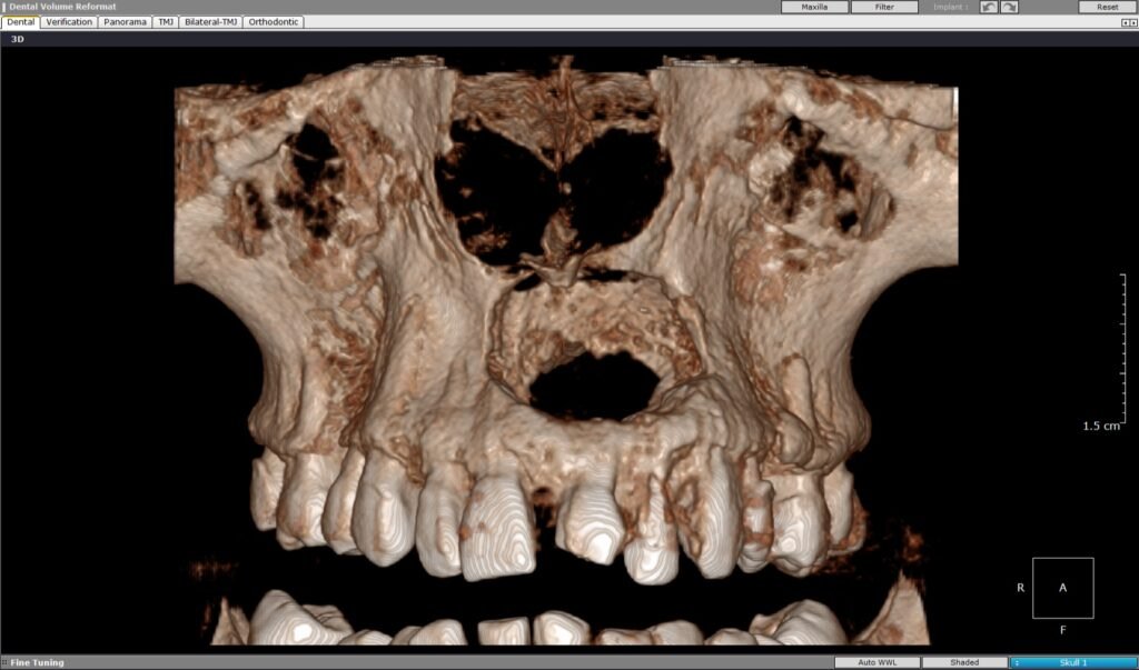
CBCT imaging offers high-resolution 3D images that provide superior visualization of oral lesions compared to traditional 2D radiographs. With CBCT, dental professionals can examine lesions from multiple angles and view them in different planes, enhancing their understanding of the lesion’s size, location, and relationship to surrounding structures.
2. Accurate Localization
CBCT allows for precise localization of lesions within the oral cavity. The detailed imaging provided by CBCT assists in determining the exact location of the lesion, including its depth, proximity to vital structures, and involvement of adjacent bone or soft tissues. This information is critical for planning surgical interventions and minimizing the risk of complications.
3. Characterization of Lesion Properties
CBCT imaging enables the characterization of lesion properties, aiding in differential diagnosis and treatment planning. The imaging can reveal important features such as the internal structure, density, and borders of the lesion. This information helps differentiate between benign and malignant lesions, cysts, infections, or developmental abnormalities, allowing for appropriate treatment decisions.
4. Assessment of Lesion Extent
CBCT provides valuable information about the extent of lesions, helping dental professionals assess the size and spread of the lesion within the oral cavity. This information is crucial for determining the appropriate treatment approach, including the need for surgical intervention, the extent of resection, or the involvement of adjacent anatomical structures.
5. Evaluation of Bony Changes
Many oral lesions can cause changes in the surrounding bone. CBCT imaging allows for a detailed evaluation of these bony changes, providing insights into the pattern, extent, and severity of bone destruction or remodeling associated with the lesion. This information is essential for treatment planning, especially in cases involving bone grafting or implant placement.
6. Treatment Planning and Follow-up
CBCT plays a vital role in treatment planning for oral lesions. The detailed imaging helps in determining the most appropriate treatment modality, whether it be surgical excision, endodontic therapy, or medical management. CBCT images also aid in the preoperative evaluation of critical anatomical structures, ensuring a safe and successful procedure. Additionally, CBCT can be used for post-treatment follow-up, allowing dental professionals to monitor healing, detect any recurrence or complications, and assess treatment outcomes.
7. Minimally Invasive Approach
CBCT imaging enables a minimally invasive approach in the diagnosis and treatment of lesions. With precise localization and accurate characterization of lesions, dental professionals can plan targeted interventions, minimizing unnecessary tissue removal and reducing patient discomfort. This not only enhances patient experience but also preserves healthy tissues and supports optimal functional and esthetic outcomes.
8. Patient Education and Communication
CBCT images are powerful visual aids for patient education and communication. The 3D nature of CBCT imaging allows dental professionals to explain the nature of the lesion, its location, and the proposed treatment plan more effectively. Patients can better understand their condition, make informed decisions, and actively participate in their treatment journey.
Conclusion
ConeBeam CT imaging has revolutionized the diagnosis and treatment planning of oral lesions. Its ability to provide high-resolution 3D images allows dental professionals to visualize and analyze lesions in detail, aiding in accurate diagnosis, treatment planning, and patient communication. With its many benefits, CBCT imaging has become an invaluable tool in modern dentistry, enhancing the quality of care and improving patient outcomes.
============================================