
A Comprehensive Guide to Fracture and Tooth Fracture Detection with ConeBeam CT
Introduction
Fractures, including tooth fractures, are common dental conditions that can have significant implications for patients’ oral health. Detecting and accurately diagnosing fractures is crucial for effective treatment planning and optimal patient outcomes. In recent years, ConeBeam Computed Tomography (CBCT) has emerged as a powerful imaging modality for the detection and evaluation of fractures. In this comprehensive guide, we will explore the role of CBCT in fracture detection, discuss its advantages, and highlight its applications in dental practice.
Understanding Fractures: A Dental Perspective
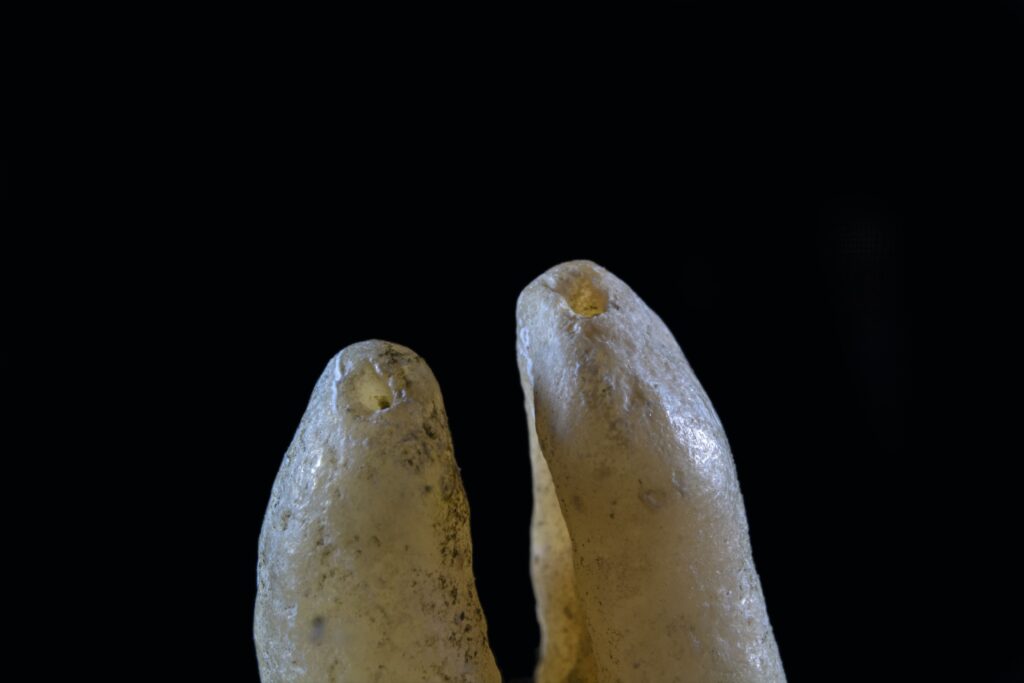
Fractures in the dental context refer to the partial or complete breakage of a tooth or the surrounding structures. They can occur due to various factors, including trauma, chewing on hard objects, bruxism (teeth grinding), or weakened tooth structure. Fractures can range from minor cracks to more severe breaks involving the enamel, dentin, and even extending into the root.
Early detection and proper diagnosis of fractures are essential to prevent further damage, preserve tooth structure, and ensure the longevity of the affected tooth. Traditionally, conventional dental radiography, such as intraoral periapical and bitewing radiographs, has been used for fracture detection. However, these 2D imaging techniques may not provide a comprehensive view of the fracture and its extent. This is where CBCT imaging proves to be invaluable.
ConeBeam CT: An Advanced Imaging Tool
ConeBeam CT is a modern imaging technique that utilizes a cone-shaped X-ray beam and a specialized detector to capture high-resolution, three-dimensional images of the oral and maxillofacial region. This imaging modality provides a detailed view of dental structures, including teeth, bone, and surrounding soft tissues, with minimal radiation exposure.
Compared to conventional radiography, CBCT offers several advantages when it comes to fracture detection:
- Enhanced Visualization: CBCT provides high-resolution 3D images, enabling a comprehensive evaluation of fractures. Dentists can visualize the fracture lines, their location, direction, and extent with greater precision.
- Accurate Diagnosis: CBCT allows for a more accurate diagnosis by providing detailed information about the fracture’s size, position, and relationship to adjacent structures. This helps dentists determine the appropriate treatment approach and predict potential complications.
- Improved Treatment Planning: With CBCT, dentists can accurately assess the fracture’s complexity and plan the most suitable treatment strategy. Whether it involves bonding, splinting, endodontic therapy, or surgical intervention, CBCT images provide valuable guidance for successful treatment outcomes.
- Evaluation of Root Fractures: Root fractures can be particularly challenging to detect using conventional radiographs alone. CBCT imaging offers enhanced visualization of root fractures, enabling early detection and appropriate management.
Applications of CBCT in Fracture Detection
CBCT imaging plays a crucial role in fracture detection across various dental specialties. Let’s explore some of the key applications of CBCT in different clinical scenarios:
1. Traumatic Injuries
CBCT is highly beneficial in cases of dental trauma, such as fractures resulting from falls, accidents, or sports-related injuries. CBCT scans provide a comprehensive assessment of both hard and soft tissues, allowing dentists to accurately diagnose fractures, identify associated injuries, and plan appropriate treatment.
2. Cracked Tooth Syndrome
Cracked Tooth Syndrome is a condition characterized by incomplete fractures that extend through the tooth structure, often causing pain upon chewing. CBCT imaging aids in the identification and localization of these elusive cracks, assisting dentists in determining the extent of the fracture and devising suitable treatment plans.
3. Vertical Root Fractures
Vertical root fractures can be challenging to diagnose due to their hidden nature. CBCT scans provide a detailed view of the root structure, allowing dentists to identify vertical fractures, determine their position, and evaluate their impact on the tooth’s prognosis. This information is crucial for deciding whether the tooth can be saved or requires extraction.
4. Endodontic Complications
Fractures can complicate endodontic treatments, affecting the success and longevity of root canal therapy. CBCT imaging helps identify fractures that may have caused treatment failure or hindered proper disinfection and obturation. By accurately diagnosing these fractures, dentists can develop appropriate retreatment plans or consider alternative treatment options.
5. Implant Planning and Placement
CBCT scans play a vital role in implant dentistry by providing a detailed evaluation of the alveolar bone and adjacent structures. In cases where fractures have resulted in tooth loss, CBCT images aid in determining the feasibility of implant placement, assessing bone quality and quantity, and identifying any underlying fractures that may affect implant stability.
6. Orthodontic Treatment
In orthodontics, CBCT imaging assists in assessing the dental and skeletal structures, identifying any underlying fractures that may impact treatment planning. It allows orthodontists to determine the optimal tooth movement and design a customized treatment plan that considers the presence of fractures for improved treatment outcomes.
Precautions and Limitations of CBCT Imaging
While CBCT imaging offers numerous advantages, it is essential to consider its limitations and take appropriate precautions:
- Radiation Exposure: Although CBCT radiation doses are significantly lower than medical CT scans, proper radiation safety measures should be followed, including the use of appropriate collimation and shielding. CBCT imaging should be justified based on the individual patient’s needs and risks.
- Artifacts: CBCT images may occasionally exhibit artifacts, such as streaking or distortion, which can affect image quality and interpretation. Awareness of these artifacts and their potential impact on fracture detection is important to ensure accurate diagnoses.
- Operator Skill and Training: CBCT imaging requires operator skill and training to obtain high-quality images and accurate interpretations. Dental professionals should undergo proper training and adhere to established protocols for image acquisition and interpretation.
- Cost Considerations: CBCT imaging may involve higher costs compared to conventional radiography. Dentists should consider the clinical necessity and potential benefits of CBCT in each case, balancing the diagnostic advantages with the associated costs.
Conclusion
ConeBeam CT imaging has revolutionized the field of fracture detection in dentistry. Its ability to provide high-resolution 3D images of dental structures offers unprecedented visualization and diagnostic accuracy. From traumatic injuries to cracked teeth and vertical root fractures, CBCT plays a vital role in identifying and evaluating fractures in various clinical scenarios. By leveraging the power of CBCT, dental professionals can enhance treatment planning, optimize patient outcomes, and deliver superior dental care.
============================================