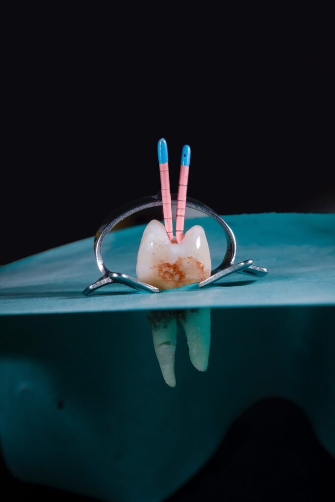
How ConeBeam CT is Revolutionizing Root Canal Evaluation
Root canal treatment is a common procedure in dentistry aimed at saving a severely damaged or infected tooth. The success of this procedure depends on accurate diagnosis and precise treatment planning. ConeBeam CT (CBCT) imaging has emerged as a revolutionary tool in root canal evaluation, providing detailed 3D images that enhance the assessment of root canal anatomy, treatment complexity, and potential complications. In this article, we will explore how CBCT is transforming the field of endodontics and revolutionizing root canal evaluation.
1. Enhanced Visualization of Root Canal Anatomy
CBCT imaging offers unparalleled visualization of root canal anatomy. Unlike traditional 2D radiographs, CBCT provides detailed 3D images that allow dental professionals to examine the tooth’s internal structures from various angles and planes. This level of visualization enables a comprehensive assessment of root canal morphology, including the number of canals, their shape, curvature, and any anatomical variations. Accurate knowledge of the root canal anatomy is crucial for successful root canal treatment, as it ensures thorough cleaning, shaping, and obturation of the entire canal system.
2. Precise Measurement and Assessment
CBCT enables precise measurement and assessment of root canal dimensions. With CBCT imaging, dental professionals can accurately measure the length, width, and curvature of the root canals. This information is invaluable for determining the appropriate instrumentation and selecting the right file sizes during the root canal procedure. It also helps in identifying potential challenges, such as narrow or calcified canals, which may require specialized techniques or instruments for successful treatment.
3. Identification of Anatomic Variations and Abnormalities
CBCT imaging allows for the identification of anatomic variations and abnormalities that may impact the success of root canal treatment. It can reveal the presence of extra canals, lateral canals, accessory canals, or root resorption, which may go undetected on 2D radiographs. By identifying these variations and abnormalities, dental professionals can tailor their treatment approach, ensuring comprehensive cleaning and disinfection of all canal spaces and improving the prognosis of the root canal procedure.
4. Detection of Pathology and Periapical Lesions
CBCT imaging is highly effective in detecting periapical lesions and pathology associated with the root canal system. It provides detailed information about the extent, size, and location of periapical lesions, such as periapical abscesses, granulomas, or cysts. This information is crucial for accurate diagnosis and treatment planning. CBCT imaging also helps in assessing the proximity of the lesion to vital structures, aiding in the identification of potential complications and guiding the appropriate treatment approach.
5. Evaluation of Treatment Outcomes
CBCT plays a significant role in evaluating the outcomes of root canal treatment. By comparing pre- and post-treatment CBCT images, dental professionals can assess the efficacy of the treatment, evaluate the quality of obturation, and identify any persistent or recurrent infections. This allows for timely intervention and retreatment if necessary, ensuring the long-term success of the root canal procedure.
6. Minimizing Treatment Risks and Complications
CBCT imaging helps in minimizing treatment risks and complications associated with root canal procedures. It provides a comprehensive understanding of the tooth’s anatomy, allowing dental professionals to anticipate challenges and plan accordingly. By identifying the proximity of important anatomical structures, such as adjacent teeth, nerves, or sinuses, CBCT helps in avoiding potential iatrogenic damage during the root canal procedure. This leads to improved patient safety and reduced treatment risks.
Conclusion
ConeBeam CT imaging has revolutionized the field of endodontics by providing detailed 3D images that enhance root canal evaluation. Its ability to visualize root canal anatomy, identify anatomic variations, detect pathology, and evaluate treatment outcomes has significantly improved the accuracy and success of root canal procedures. With CBCT, dental professionals can provide patients with more precise and effective root canal treatment, preserving natural teeth and improving oral health outcomes.
============================================

